International Journal of Dermatology and Clinical Research
Non-Small Cell Lung Cancer-A Case of the Bazex
Timothy Cook* and Carles Escriu
The Clatterbridge Cancer Centre, Liverpool L78YA, UK
Cite this as
Cook T, Escriu C. Non-Small Cell Lung Cancer-A Case of the Bazex. Int J Dermatol Clin Res. 2024;10(1):001-004. Available from: 10.17352/2455-8605.000050Copyright
© 2024 Cook T, et al. This is an open-access article distributed under the terms of the Creative Commons Attribution License, which permits unrestricted use, distribution, and reproduction in any medium, provided the original author and source are credited.Bazex syndrome is a rare form of paraneoplastic syndrome affecting the skin, rarely identified as an underlying cancer cause at presentation. The current case highlights, as well as a literature review of cases, a 76-year-old man with a smoking history and no skin medical history who had a delayed diagnosis of Non-Small Cell Lung Cancer (NSCLC). Initial pre-diagnosis skin plaque treatment failed, with progressing shortness of breath leading to a finding of lung cancer. Plaques were classical in this case and did respond to the chemotherapy as previously reported, but not previously noted with the combination used in this case. Although the skin plaques responded to treatment, the patient did eventually die of advancing lung cancer.
Introduction
Bazex syndrome (BAK) is a paraneoplastic psoriasiform dermatitis, predominately seen in Squamous Cell Carcinomas (SCC) of aero-digestive tract and non-small cell lung cancers [1,2]. Lesions are seen mainly in adult males (>60 years old (YO)) >3:1 women, with an alcohol and smoking history noted in 28% of cases noted to be alcohol and smoking [1,2].
Clinically, lesions may be erythematous, scaling, and appear psoriatic-like, which may lead to a delayed diagnosis of cancer while benign skin causes are treated [2]. Acral lesions may be considered pathognomonic of BAK upon confirmation of carcinoma with common distribution of lesions on: ears (79% & 28%), nails (75% & 57%), nose (63% & 33%), fingers (61% & 29%), hands (57% & 38%), feet (50% & 30%) and less commonly the trunk (13% & n/a), scalp (7% & n/a), elbows (19% & n/a) and cheeks (19% & n/a) [1,2].
Treatment of the underlying cancer has been found to successfully treat the skin lesions when therapy is established with cases noting response to NSCLC BAK syndrome lesions after chemotherapy, immunotherapy, and surgical intervention (see figure 2) [3-6].
The current case reports BAK syndrome in a 76-year-old male following a delayed diagnosis and details his diagnosis and treatment journey to date while he remains on current active first-line management.
Within the report is a brief review of the lung cancer cases and BAK syndrome, showing literature available via Medline and Pubmed from 2011-2021 in the English language using the search term ((Bazex) OR (acrokeratosis)) AND (TitleCombined:(lung cancer OR carcinoma)).
Consent
Consent was obtained to use images about this report, identifiers are anonymized and no patient personal information is used within this article.
Timeline
December 2020 – January 2023.
Patient presentation
76-year-old retired white British male presented with a 6-month history of psoriatic-like plaques on fingers and toes bi-laterally, progressing to the scalp and torso, with >10% weight loss and progressing breathlessness two weeks before diagnosis.
He has a > 40-pack-year smoking history and a past medical history of Chronic Obstructive Pulmonary Disease (COPD) and hypertension, taking medicines; amlodipine 5 mg, acitretin 20 mg, atorvastatin 10 mg nocte, furosemide 40 mg mane, aspirin 75 mg, tiotropium 10 mcg mane and salbutamol inhaled 100 mcg/dose, with no known drug allergies.
No family history of Bazex acrokeratosis neoplastica syndrome or lung cancer was present.
Delayed diagnosis
Initial treatment for scaling dermatosis was initiated by the community doctor with dermol 500 and dermovate cream, which gained no symptomatic benefit over 6 months March – September, later given acitetin 20 mg with no continued benefit. New shortness of breath triggered a chest x-ray via the community GP which highlighted a supraclavicular mass and a right hilar mass, leading to a two-week wait for suspected cancer.
Diagnostics
A Computer Tomography (CT) and a positron emission tomography CT scan (PET-CT) confirmed stage 3b lung cancer, with a 7 cm mass in the right lower lobe with atelectasis and right upper lobe nodal lesion with a 4 cm supraclavicular lesion and a paratracheal node at 3.5 cm, confirmed as avid on a positron CT scan. The supraclavicular node was biopsied showing a squamous cell carcinoma with a programmed death ligand -1 as 10% and no actionable mutations, next-generation sequencing was not performed.
Stage:
T4N3M0 – 3b [7].
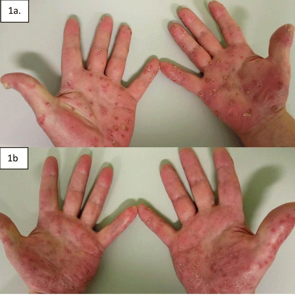
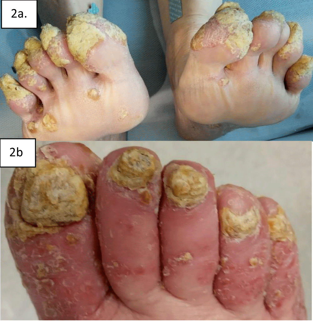
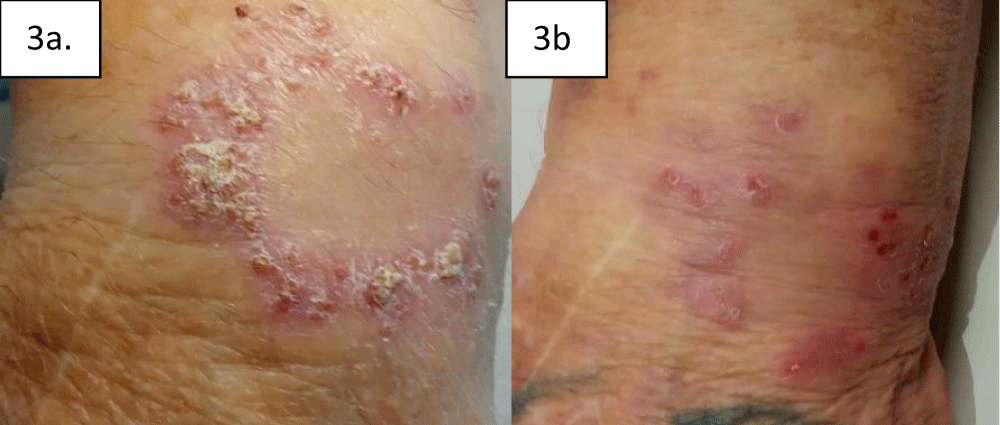
Figures 1-3: Acral and wrist lesions (1a, 2a, & 3a) Photographed before treatment in December 2020 and April 2021 (1b, 2b & 3b), after 6 cycles of Carboplatin, paclitaxel and pembrolizumab.
Examination
The examination took place before cycle one of chemo/immunotherapy, or any anti-cancer treatment on the same day. A comparison of treatment will be detailed in the discussion.
Overall the patient was comfortable at rest, with no mobility aids, no oxygen, and no indwelling catheters or lines in situ. The end-of-bed assessment identified plaques upon fingernails bi-laterally.
Lung exam: No additional berthing sounds were identified, with good air expansion. Saturation was 96% on room air with a respiration rate of 16 breaths per minute at rest.
Heart exam: No additional sounds or murmurs were identified, with a pulse rate of 58 beats per minute and a blood pressure of 103/53.
Abdominal exam: A single plaque was identified in the epigastric area, no scars were noted, with no masses, a soft abdomen on palpation, and bowel sounds throughout.
Hands: Plaques (pre-treatment) noted on fingernails bi-laterally, crusting and yellow with thickened perioncychium and mild palm erythema bi-laterally.
Toenails: Equally to fingers, yellow scaling plaques bi-laterally were noted with erythematous cracks on the heels bi-laterally.
Other: Three lesions were noted on the scalp and one on the post oracular on the left side.
Treatment
Triple therapy was initialized based on squamous metastatic NSCLC with a PDL-1 of 10%, given Carboplatin, Paclitaxel, and Pembrolizumab was administered in four cycles, following which, Pembrolizumab immunotherapy continued as a single agent treatment [8]. Radiotherapy 20 grey in 5 fractions was given to reduce the paratracheal mass, with continued Pembrolizumab to 10 cycles before progressive disease n mediastinum and superior vena cava obstruction, before the patient died of progressive disease.
Scans performed every 3 months measured with iRECIST criteria [9], with the best outcome Partial Response (PR) to treatment and an overall time to progression of 10 months from the start of treatment.
Discussion
Delayed diagnosis of BAK syndrome may prolong ant-cancer treatment leading to diagnosis at a later stage, limiting treatment options for patients, which parallels the current case who waited 6 months from the onset of plaque lesions to diagnosis of lung cancer [1]. Zarzour, Singh, Andea & Cafardi (2011) noted the death of a small cell lung cancer patient 6 months following diagnosis despite a 2-year history of a hyperpigmented rash over forearms and legs, diagnosed as BAK syndrome [1,2,10]. Similarly, an Epidermal Growth Factor Receptor mutation (EGFR) was diagnosed with metastatic bone lesions after a 5 year history of pruritic, acral lesions, suggesting earlier recognition may have allowed lower diagnostic stage [6].
The current case noted a 6-month delay pre-treatment, leading to a diagnosis of 3b NSCLC with supraclavicular lymph node spread, meaning an advanced stage of lung cancer untreatable to radical surgery or radiation, giving a poor prognosis with systemic treatment, although the current treatment regime of chemotherapy combined immunotherapy may lead to prolonged survival [7,8].
Psoriatic lesions occurred in the current case on the hands, feet, nose, nails, auricular helices, and scalp in the current case, with a lesion on the trunk suggesting advanced carcinoma correlation [11,12]. Typically the lesions will respond (Table 1) to anti-cancer therapy as was the case here, the individual who failed to respond died within 6 months of diagnosis and unusually had SCLC [10].
Previous cases have shown symptomatic benefits from adjuvant treatment with acitretin, used to treat severe psoriasis, at 0.25-1mg/kg body weight [11,13].
The current case has shown how Pembrolizumab combined with chemotherapy therapy in advanced/metastatic NSCLC [8], may reduce plaques in BAK syndrome (Figure 1 A-a B-b) which is paralleled by [4], which showed an improvement in the distal extremities after 7 cycles of Pembrolizumab.
Pathophysiology of BAK syndrome may be related to the cross-reactivity of tumor antigens irritating the skin, growth factors associated with epidermal growth factor, and vitamin deficiency such as Zinc and vitamin A [1,12]. Amanon, et al. (2016) Showed serum immunoglobulin e and eosinophil count elevated in an 82 YO SCC of the lung, with reduced levels after tumor removal, suggesting a role of the Th2-type cytokine response leading to an elevated inflammation, which may cross-react causing keratin-based skin plaques seen in BAK syndrome, although the reactive response to subcutaneous cancer itself could not be ruled out as well as raised inflammation as a result of patients positive smoking status [3].
The current case survived approximately 12 months post-treatment with chemotherapy and pembrolizumab, compared to 15.9 months in the literature for NSCLC SCC patients [8].
Diagnosis in the current case was on observation capacity, failure to respond to anti-psoriatic medication, and response to anti-cancer therapy, although no biopsy may be a limitation here [1]. Further limitations are a lack of tracked biomarkers throughout treatment, with immunoglobulin E, also not detailed, as suggested to be raised in BAK syndrome by Amanon, et al. [3].
In summary, the current case highlights that BAK syndrome in SCC NSCLC can be successfully treated with combined chemotherapy and immunotherapy, with lesion/plaque regression corresponding to successful anti-cancer treatment, although the patient did die of advanced NSCLC. Plaques in BAK syndrome are not commonly responsive to psoriatic therapy, and a smoker with a resistant history of psoriatic lesion treatment should be considered for an upper digestive or lung cancer diagnosis to rule out an underlying malignancy [1].
Ethics approval
Ethical guidelines were followed, with written consent obtained for images.
De-identification
All information remains anonymized.
- Bolognia JL, Brewer YP, Cooper DL. Bazex syndrome (acrokeratosis paraneoplastica): an analytic review. Medicine. 1991;70(4):269-280. Available from: https://journals.lww.com/md-journal/citation/1991/07000/bazex_syndrome__acrokeratosis_paraneoplastica__an.4.aspx
- Räßler F, Goetze S, Elsner P. Acrokeratosis paraneoplastica (Bazex syndrome) - a systematic review on risk factors, diagnosis, prognosis and management. J Eur Acad Dermatol Venereol. 2017;31(7):1119-1136. Available from: https://doi.org/10.1111/jdv.14199
- Amano M, Hanafusa T, Chikazawa S, Ueno M, Namiki T, Igawa K, et al. Bazex syndrome in lung squamous cell carcinoma: high expression of epidermal growth factor receptor in lesional keratinocytes with Th2 immune shift. Case Rep Dermatol. 2016;8(3):358-362. Available from: https://doi.org/10.1159/000452827
- Aoshima Y, Karayama M, Sagisaka S, Yasui H, Hozumi H, Suzuki Y, et al. Synchronous occurrence of Bazex syndrome and remitting seronegative symmetrical synovitis with pitting edema syndrome in a patient with lung cancer. Intern Med. 2019;58(22):3267-3271. Available from: https://doi.org/10.2169/internalmedicine.3032-19
- Mititelu R, Powell M. A case report of resolution of acrokeratosis paraneoplastica (Bazex syndrome) post resection of non-small-cell lung carcinoma. SAGE Open Med Case Rep. 2019;7:2050313X19881595. Available from: https://doi.org/10.1177/2050313x19881595
- Zhao J, Zhang X, Chen Z, Wu JH. Case report: Bazex syndrome associated with pulmonary adenocarcinoma. Medicine (Baltimore). 2016;95(2):e2415. Available from: https://doi.org/10.1097/md.0000000000002415
- Matilla JM, Zabaleta M, Martínez-Téllez E, Abal J, Rodríguez-Fuster A, Hernández-Hernández J. New TNM staging in lung cancer (8th edition) and future perspectives. J Clin Transl Res. 2020;6(4):145-154. Available from: https://pmc.ncbi.nlm.nih.gov/articles/PMC7837738/
- Paz-Ares L, Luft A, Vicente D, Tafreshi A, Gümüş M, Mazières J, et al. Pembrolizumab plus chemotherapy for squamous non-small-cell lung cancer. N Engl J Med. 2018;379(21):2040-2051. Available from: https://doi.org/10.1056/nejmoa1810865
- Persigehl T, Lennartz S, Schwartz LH. iRECIST: how to do it. Cancer Imaging. 2020;20(1):2. Available from: https://doi.org/10.1186/s40644-019-0281-x
- Zarzour JG, Singh S, Andea A, Cafardi JA. Acrokeratosis paraneoplastica (Bazex syndrome): report of a case associated with small cell lung carcinoma and review of the literature. J Radiol Case Rep. 2011;5(7):1-6. Available from: https://doi.org/10.3941/jrcr.v5i7.663
- Bazex A, Griffiths A. Acrokeratosis paraneoplastica--a new cutaneous marker of malignancy. Br J Dermatol. 1980;103(3):301-6. Available from: https://doi.org/10.1111/j.1365-2133.1980.tb07248.x
- Rodrigues IA Jr, Gresta LT, Cruz RC, Carvalho GG, Moreira MH. Bazex syndrome. An Bras Dermatol. 2013;88(6 Suppl 1):209-11. Available from: https://doi.org/10.1590/abd1806-4841.20132488
- Vaynshtok PM, Tian F, Kaffenberger BH. Acitretin amelioration of acrokeratosis paraneoplastica (Bazex syndrome) in cases of incurable squamous cell carcinoma of the hypopharynx. Dermatol Online J. 2016;22(9):13030/qt8n96v45x. Available from: https://pubmed.ncbi.nlm.nih.gov/28329620/
Article Alerts
Subscribe to our articles alerts and stay tuned.
 This work is licensed under a Creative Commons Attribution 4.0 International License.


 Save to Mendeley
