Global Journal of Perioperative Medicine
Primary pulmonary myxoid sarcoma: A case report and review of the literature
1Department of Surgery, Henry Ford Health, Detroit, Michigan, USA
2Department of Pathology, Henry Ford Health, Detroit, Michigan, USA
3School of Medicine, University of Texas Medical Branch, Galveston, Texas, USA
Author and article information
Cite this as
Hutchings H, Schwarze E, Ahsan B, Cox J, Okereke I (2023) Primary pulmonary myxoid sarcoma: A case report and review of the literature. Glob J Perioperative Med. 2023; 7(1): 001-003. Available from: 10.17352/gjpm.000012
Copyright License
© 2023 Hutchings H, et al. This is an open-access article distributed under the terms of the Creative Commons Attribution License, which permits unrestricted use, distribution, and reproduction in any medium, provided the original author and source are credited.Background: Primary pulmonary myxoid sarcoma is a very rare tumor, with less than 25 cases being described in the literature. When found in the bronchial tree, an R0 resection with post-operative follow-up is sufficient for curative treatment. We present a case of primary pulmonary myxoid sarcoma in a middle-aged man with an associated review of the literature to highlight this rare tumor.
Case presentation: A 62-year-old man with a 40-pack-year smoking history presented to the thoracic surgery clinic for evaluation of a suspicious lung nodule. Pre-operative testing revealed a spiculated mass in the left upper lobe and the biopsy was non-diagnostic. The patient was taken for left upper lobectomy tri-segmentectomy with mediastinal lymph node dissection. Final pathology of the nodule revealed primary pulmonary myxoid sarcoma with an Ewing sarcoma breakpoint region 1 on chromosome 22 with activating transcription factor 1 (EWSR-ATF1) fusion transcript consistent with the diagnosis.
Conclusion: Primary pulmonary myxoid sarcoma is an exceedingly rare sarcoma of the lung with less than 25 reported cases in the literature. This case report highlights our experience treating primary pulmonary myxoid sarcoma in a middle-aged man, and a review of current treatment recommendations in the available published literature.
PPMS: Primary Pulmonary Myxoid Sarcoma; AFH: Angiomatoid Fibrous Histiocytoma; EWSR-ATF1: Ewing Sarcoma Breakpoint Region 1 on chromosome 22 with activating transcription Factor 1
Introduction
Primary Pulmonary Myxoid Sarcoma (PPMS) is an exceedingly rare tumor that can mimic Angiomatoid Fibrous Histiocytoma (AFH) [1]. PPMS tends to be predominantly myxoid cellularity with spindle, stellate, or polygonal cells as compared to AFH, and although anatomic variability exists, the majority of PPMS tumors are comprised of endobronchial components. We describe our experience of a case of PPMS in a middle-aged patient and present available data on PPMS in the literature. The Institutional Review Board (IRB) waived IRB approval as this study was considered exempt from IRB approval given the single case nature. The subject provided informed written consent for the publication of the study data.
Case presentation
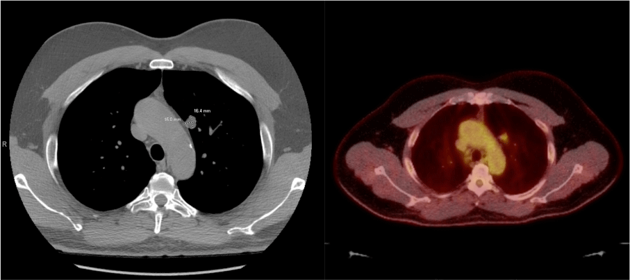
Our patient was a 62-year-old man with a 40-pack-year smoking history who was referred for evaluation of a suspicious lung nodule. A Computed Tomography (CT) scan revealed a spiculated mass measuring 1.8 centimeters (cm) in the left upper lobe (Figure 1). Percutaneous biopsy was non-diagnostic. An integrated whole-body positron emission tomography (PET)-CT scan showed the mass to have a standard uptake value of 3.2 (Figure 1). No mediastinal activity was noted on the PET-CT scan.
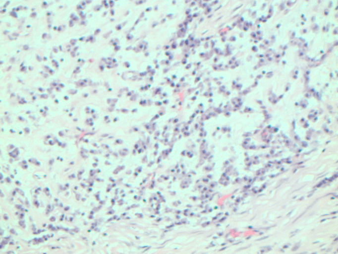
He underwent a Video-Assisted Thoracoscopic (VATS) left upper lobectomy tri-segmentectomy with mediastinal lymph node dissection. The final pathologic review revealed a low-grade spindle cell lesion measuring 1.8 x 1.6 x 1.3 cm. Greater than 90 percent of the tumor was composed predominantly of a myxoid component, leading to a diagnosis of PPMS (Figure 2). The tumor was negative for the following immunostains: CKAE1/AE3, smooth muscle actin, calponin, S100, and CD34. The tumor contained an Ewing sarcoma breakpoint region 1 on chromosome 22 with activating transcription factor 1 (EWSR-ATF1) fusion transcript consistent with PPMS [2]. All lymph nodes were negative for malignancy. The final pathological stage was pT1bN0M0. He had an uneventful postoperative course and was discharged to home in good condition. On follow-up 12 months after surgery, the patient was in good health with no evidence of recurrent disease.
Discussion
PPMS is an exceedingly rare tumor, with only 24 previously reported cases in the literature. Histologically the tumor mimics AFH but contains an overwhelmingly predominant myxoid component, lacks prominent lymphoid rimming, and typically forms anastomosing cords that resemble an overall lacy pattern [1,3]. EWSR fusion proteins are a class of oncogenes characterized by fusions of the prion-like disordered N terminal domain. The presence of an EWSR-ATF1 fusion transcript with a predominant myxoid component generally confirms the diagnosis of PPMS [4]. PPMS tumors have been reported to occur in patients of a wide age range [3]. Although rare, the prognosis is similar to other non-small cell lung cancers based on TNM categorization.
In reviewing the literature, the mean age for this diagnosis is 48.4 years old, with an even distribution between men and women (Supplemental Table 1) [1,3]. Clinical presentation of PPMS tumors can vary, with some incidentally found on radiographic imaging in other patients with active symptomology, ranging from unintentional weight loss to active respiratory manifestations of hemoptysis and dyspnea [5]. Genetic heterogeneity exists within these rare tumors. Currently, there are only two prior recorded cases of PPMS tumors presenting with the EWSR-ATF1 fusion gene. More often patients present with an EWSR-CREB1 fusion gene. The EWSR fusion protein codes for a nuclear protein involved in cell meiotic division, maintenance of DNA and microtubules, and other functions. The fusion of an activating transcription factor (CREB1, ATF1, and CREM) allows for activation of this gene and dysregulation of the cell’s meiotic activity [3].
Treatment
Most cases of PPMS with the EWSR fusion gene have a favorable prognosis [1]. Most patients present with an asymptomatic mass. Treatment for these cancers is complete R0 surgical resection with postoperative surveillance [5]. Early diagnosis, complete resection, and routine postoperative surveillance are important to increase the chances of cure. Most tumors follow an indolent course, but there are some cases that demonstrate recurrence of disease necessitating close follow-up. The existence of the fusion transcripts does not appear to alter prognosis.
Conclusion
In conclusion, PPMS is an exceedingly rare, low-grade sarcoma of the lung that often offers a favorable prognosis with complete surgical resection. Due to the sparse literature regarding this diagnosis, further work is needed to identify larger case series and develop more specific treatment guidelines for PPMS.
Ethics statement
The Institutional Review Board (IRB) waived IRB approval as this study was considered exempt from IRB approval given the single case nature. The subject provided informed written consent for the publication of the study data.
Consent for publication
The subject provided written informed consent for publication of this case report.
Author contributions
IO, BA: treatment of the patient, pathologic diagnosis; HH, ES, JC: Manuscript writing and literature review. All authors: Review and approval of final manuscript.
- Gui H, Sussman RT, Jian B, Brooks JS, Zhang PJL. Primary Pulmonary Myxoid Sarcoma and Myxoid Angiomatoid Fibrous Histiocytoma: A Unifying Continuum with Shared and Distinct Features. Am J Surg Pathol. 2020 Nov;44(11):1535-1540. doi: 10.1097/PAS.0000000000001548. PMID: 32773530.
- Metzgeroth G, Ströbel P, Baumbusch T, Reiter A, Hastka J. Hepatoid adenocarcinoma - review of the literature illustrated by a rare case originating in the peritoneal cavity. Onkologie. 2010;33(5):263-9. doi: 10.1159/000305717. Epub 2010 Apr 13. PMID: 20502062.
- Prieto-Granada CN, Ganim RB, Zhang L, Antonescu C, Mueller J. Primary Pulmonary Myxoid Sarcoma: A Newly Described Entity-Report of a Case and Review of the Literature. Int J Surg Pathol. 2017 Sep;25(6):518-525. doi: 10.1177/1066896917706413. Epub 2017 Apr 28. PMID: 28449608.
- Wang WL, Mayordomo E, Zhang W, Hernandez VS, Tuvin D, Garcia L, Lev DC, Lazar AJ, López-Terrada D. Detection and characterization of EWSR1/ATF1 and EWSR1/CREB1 chimeric transcripts in clear cell sarcoma (melanoma of soft parts). Mod Pathol. 2009 Sep;22(9):1201-9. doi: 10.1038/modpathol.2009.85. Epub 2009 Jun 26. PMID: 19561568.
- Nishimura T, Ii T, Inamori O, Konishi E, Yoshida A. Primary Pulmonary Myxoid Sarcoma with EWSR1::ATF1 Fusion: A Case Report. Int J Surg Pathol. 2023 Feb;31(1):88-91. doi: 10.1177/10668969221095457. Epub 2022 Apr 24. PMID: 35466725.
Article Alerts
Subscribe to our articles alerts and stay tuned.
 This work is licensed under a Creative Commons Attribution 4.0 International License.


 Save to Mendeley
