Global Journal of Perioperative Medicine
Identification of pre-operative, intra-operative and post-operative risk factors for aspiration pneumonia in patients undergoing abdominal surgery
Kyle John Lindfield1* and Andrew Little2
2Gold Coast University Hospital, Australia
Cite this as
Lindfield KJ, Little A (2019) Identification of pre-operative, intra-operative and post-operative risk factors for aspiration pneumonia in patients undergoing abdominal surgery. Glob J Perioperative Med 3(1): 001-006. DOI: 10.17352/gjpm.000006Objective: To investigate the pre-operative, intra-operative and post-operative risk factors associated with aspiration pneumonia in patients undergoing abdominal surgery. We also aimed to identify the risk-factors that were associated with increased mortality.
Design: Retrospective audit.
Setting: Single regional centre located in Australia.
Participants: Patients that were admitted under the general surgery team at a regional hospital in Australia were reviewed to confirm the presence of aspiration pneumonia as a complication during their admission. A total of 19 patients were identified that had a confirmed diagnosis of aspiration pneumonia between December 2013 and November 2016. The medical record of each case of aspiration pneumonia was reviewed in order to identify high-risk features for the development of aspiration pneumonia.
Results: The incidence of aspiration pneumonia was found to be 0.3% (19/6431 presentations) between December 2013 and November 2016. The procedure associated with the highest risk of developing aspiration pneumonia was laparoscopic surgery for division of adhesions, in which aspiration pneumonia occurred in 3 of 127 cases (2.3%).
Patients in the non-survivor group were older than the survivor group (81 +/- 12.0 vs 72 +/- 9.9) and had higher American Society of Anaesthesiologists (ASA) physical status score (3.7 +/- 0.6 vs 2.6 +/- 0.6). A history of pre-existing neurological disorders and gastro-oesophageal reflux disorder (GORD) were the most common risk factors for aspiration pneumonia identified. Both of these conditions were present in a total of 8 (42%) patients. Emergency surgical procedures accounted for 14 (74%) of patients that developed aspiration pneumonia in the perioperative setting.
Conclusion: There is a low overall incidence of aspiration pneumonia in patients admitted for gastrointestinal surgery or emergency endoscopy (0.3%). Aspiration with severe consequences tended to occur in patients who were elderly (age > 70-years) and had an ASA physical status score of 3 or more. Pre-existing neurological deficit and GORD were the most common risk factors for the development of aspiration pneumonia. Our study supports the use of a screening tool for the pre-operative identification of patients at risk of pulmonary aspiration. We recommend the implementation of a protocol for managing high-risk patients in the perioperative setting, which includes consideration of the following factors: 1) Prescription of a Proton Pump Inhibitor (PPI) or a Histamine Receptor (H2-R) Antagonist on admission; 2) Implementation of an opioid and sedative sparing technique in the perioperative setting; 3) Consideration of early nasogastric tube insertion, with reinsertion if it is dislodged; 4) Nursing in a 30-degree position with the head up; 5) White board communication tool at the bedside to communicate important dietary information; 6) Multidisciplinary team involvement with speech pathology and physiotherapy input; 7) Speech pathology review prior to eating in the post-operative period if the patient is considered high-risk.
Introduction
Background
Aspiration pneumonia is a rare, but significant complication that contributes to the overall morbidity and mortality of patients admitted to hospital for gastrointestinal surgery. The incidence of aspiration pneumonia is reported to be approximately 1% and the associated mortality is reported to be as high as 70% [1,2]. Furthermore, aspiration pneumonia is associated with a substantial economic burden on the healthcare system due to increased requirement for Intensive Care Unit (ICU) admission and prolonged hospital length of stay [1].
Patients undergoing abdominal surgery have an increased risk of aspiration pneumonia [3]. A major determinant of pulmonary aspiration is the development of bowel ileus in the postoperative period following abdominal surgery [4]. Other factors that increase the risk of aspiration pneumonia include: altered level of consciousness; pre-existing neurological conditions; gastro-oesophageal reflux disorder (GORD); elderly age; obesity; hiatus hernia; and oesophageal dysmotility syndrome [4]. In contrast, patient position with 30° elevation of the upper body has been demonstrated to be a protective factor against the development of pulmonary aspiration in the ICU [5].
This retrospective study aims to investigate the pre-operative, intra-operative and post-operative risk factors associated with aspiration pneumonia in patients undergoing abdominal surgery. The early identification and management of patients at risk of pulmonary aspiration may help to reduce morbidity and mortality in the perioperative setting. This study also plans to develop a simple checklist which can assist clinicians to identify patients at risk of aspiration pneumonia and guide appropriate management strategies to reduce its incidence.
Method
Method of evaluation: This study is a retrospective audit of patients admitted under the General Surgery team at a regional hospital in Australia between December 2013 to November 2016. Patients were included in the study if they had objective evidence of aspiration pneumonia during their admission for gastrointestinal surgery or emergency endoscopy. Patients with pre-operative, intra-operative and post-operative aspiration pneumonia were included in this study. At the time of study completion, there was no guideline in place to support clinicians in the identification of patients at risk of aspiration pneumonia.
Eligible patients were identified by searching the patient database on the Business Objects reporting tool using the International Statistical Classification of Diseases and Related Health Problems, Tenth Edition, Australian Modification (ICD-10-AM) code for aspiration pneumonia. The electronic and paper medical records of identified patients was reviewed by a single investigator. The presence of aspiration pneumonia was confirmed in all cases with either: 1) Objective clinical evidence of pulmonary aspiration (crepitations, tachypnoea, tachycardia, fever, hypoxaemia); or 2) Objective radiological evidence (plain film chest radiography or computed tomography scan). Patients were excluded if they did not undergo surgical intervention during their admission.
Patient demographics were collected and recorded on an electronic spreadsheet (Microsoft Excel 2016, Microsoft Corporation). The variables that were recorded in the electronic spreadsheet for each patient are presented in table 1. pre-operative variables recorded were: age, gender, American Society of Anaesthesiologists (ASA) physical status score, pre-operative insertion of nasogastric tube, medications administered pre-operatively, and patient risk factors for aspiration (pre-existing neurological disorder, GORD, obesity, diabetes, renal impairment, sepsis and oesophageal dysmotility). intra-operative variables recorded were: surgical risk factors for aspiration (patient positioning, duration of surgery, experience of surgeon, laparoscopic surgery, ileostomy/colostomy, red cell transfusion) and anaesthetic risk factors (experience of anaesthetist, induction method, type of anaesthetic and amount of opioid administered). post-operative variables included were: day of post-operative pulmonary aspiration, days in the ICU, total hospital length of stay, requirement for mechanical ventilation, nursing 30 degrees upright, need for intubation, nasogastric tube post-operatively, time to first bowel motion, time to feeding, type of diet and prescription of post-operative opioids.
The sample size of this study was limited by the absolute number of patients that met the inclusion criterion within the hospital. We included all patients who were admitted under the General Surgery team that underwent either abdominal surgery or emergency endoscopy with a confirmed diagnosis of aspiration pneumonia either pre-operatively, intra-operatively or post-operatively.
Analysis
Continuous variables are presented as mean +/- standard deviation (SD). Categorical variables are presented as number (n) and percentages (%). Risk factors for increased mortality associated with aspiration were identified by comparing all recorded parameters between survivors and non-survivors. Given that the total patient population was small (n = 19), particularly in the non-survivor group (n = 3), the power of this study was limited as there were insufficient numbers to infer statistically significant differences between the two study groups.
Ethical Issues
Being a retrospective data audit of de-identified patient records, requirements for exemption from ethical review in accordance with section 5.1.22 of the National Statement on Ethical Conduct in Human Research were met, by being a negligible risk activity using de-identified data. These conditions, as outlined in Appendix 1, were adhered to.
Results
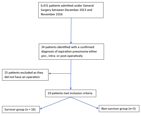
A total of 6,431 patients were admitted under the General Surgery team between December 2013 and November 2016 (Figure 1). A diagnosis of aspiration pneumonia was confirmed in 34 of these patients. Fifteen patients were excluded from the study because they did not receive surgical intervention during their admission. The remaining 19 patients underwent either abdominal surgery or emergency endoscopy and were included in this study. The following surgical interventions were performed: 6 laparotomy/laparoscopic bowel resections, 6 emergency gastroscopies, 3 division of adhesion procedures, 3 hernia repair procedures and 1 cholecystectomy. Fourteen (74%) of these procedures were performed under emergency conditions.
The timing and cause of aspiration pneumonia was reviewed in each case. Three patients aspirated in the Emergency Department, prior to being admitted to the ward (1 patient was known to Palliative Care with an oesophageal stricture, 1 patient had profound nausea and vomiting in the Emergency Department and 1 patient aspirated after receiving 5mg droperidol for agitation). Four patients aspirated intra-operatively (1 during an awake fibre-optic intubation, 1 was reported by the Anaesthetist to have a soiled airway at induction, 1 aspirated during a gastroscopy for a food bolus without an endotracheal tube, and 1 aspirated during extubation following a case of severe laryngospasm). The remaining 12 patients aspirated post-operatively on the surgical ward.
Incidence and outcome of aspiration pneumonia
The incidence of aspiration pneumonia according to the type of surgical procedure is displayed in table 2. The procedure associated with the highest risk of developing aspiration pneumonia was laparoscopic surgery for division of adhesions, in which aspiration pneumonia occurred in 3 of 127 cases (2.3%). Following this, bowel resection (either via laparotomy or laparoscopy) resulted in aspiration pneumonia in 6 of 425 cases (1.4%).
In patients who aspirated in the post-operative period, aspiration occurred at mean post-operative day 5.0 +/- 4.3. In survivors it occurred on day 4.18 +/- 2.6 and in non-survivors on day 9.5 +/- 7.6. Nine patients (47%) were admitted to the ICU, with an average ICU length of stay of 5.8 +/- 3.1 days. Two (11%) patients were intubated prior to their admission to ICU, one patient was reintubated at the end of the surgical procedure due to severe laryngospasm and the other patient remained intubated following the operation. The average hospital length of stay for the entire group was 20.2 +/- 17.4 days. The overall mortality was 16% (3 of 19 cases).
Risk factors for mortality
The pre-operative variables that were recorded in this study are displayed in table 3. A comparison is made between survivors and non-survivors. Patients in the non-survivor group were older than the survivor group (81 +/- 12.0 vs 72 +/- 9.9) and had higher ASA score (3.7 +/- 0.6 vs 2.6 +/- 0.6). A history of pre-existing neurological disorders and GORD were the most common risk factors for aspiration pneumonia, each being present in 8 of 19 (42%) patients. The insertion of a nasogastric tube in the pre-operative period was performed in only 7 of 19 (37%) patients.
The intra-operative variables recorded in this study are displayed in table 4. A longer duration of surgical procedure was found in the non-survivor group (128.0 +/- 59.2 minutes) compared to the survivor group (116.4 +/- 66.1 minutes). All procedures were performed by a Consultant Surgeon and a Consultant Anaesthetist. Fourteen procedures (74%) were performed under emergency conditions. Only 6 (42%) of these emergency cases were documented as a Rapid Sequence Induction (RSI), however, a subsequent 5 cases documented an RSI dose of Rocuronium or Suxamethonium and thus RSI could be inferred in 11/14 (79%) cases.
The post-operative variables that were recorded in this study are displayed in table 5. There was a trend towards the non-survivor group aspirating at a later postoperative day (9.5 +/- 7.6) compared to the survivor group (5.7 +/- 2.6). There were no ICU admissions for the non-survivor group whereas there were 9 (56%) ICU admissions in the survivor group with a mean length of ICU stay of 5.8 +/- 3.1 days. Opioid consumption was calculated as the mean amount of opioid (oral morphine equivalent) consumed per day within the first 10 days of admission with the non-survivors (22+/-19mg/day) tending to consume more opioid on average compared to the survivors (16+/-12mg/day).
Twelve (63%) patients received a nasogastric tube (NGT) during their admission, with 9 (75%) of these patients having it inserted prior to the aspiration event as a form of prophylaxis. There was no difference between the two groups in relation to the first day of feeding which commenced on average postoperative day 1.8 +/- 1.4. The mean time to first bowel motion was on postoperative day 2.8 +/- 2. A total of 2 patients required mechanical ventilation in ICU and another 2 patients required Non-Invasive Ventilation (NIV) with Bi-PAP or CPAP. The remaining 15 patients required either high flow nasal prongs (HFNP), low flow nasal prongs (LFNP) or no oxygen therapy. Antibiotics were commenced in all patients, bronchodilators were administered in 9 (47%) patients, steroids were administered in 3 (16%) patients and inotropes were required in 2 (11%) patients.
Discussion
Between December 2013 and November 2016, a total of 6,431 patients were admitted under the General Surgery team at a regional hospital in Australia. Of the 6,431 admitted patients, only 19 patients (0.3%) developed aspiration pneumonia. The incidence of aspiration pneumonia (0.3%) calculated in this study was below the incidence of 1.0% reported in other studies [1]. Aspiration pneumonia is a rare, but clinically relevant event due to the significant morbidity and mortality associated with its occurrence. Previous studies have reported a mortality rate of up to 70% [2]. We report a mortality rate of 16% in patients undergoing gastrointestinal surgery or emergency endoscopy. The reason for this observed difference in incidence and mortality can be explained by the different patient groups that were included in previous studies. Our study focuses exclusively on patients admitted for emergency endoscopy or gastrointestinal surgery, whereas previous studies have included a wide range of both surgical and non-surgical patient groups [1,2].
Risk factors for mortality have been documented in the literature [3]. In this study, we aimed to define the characteristics of patients that were admitted to hospital for abdominal surgery and who went on to develop aspiration pneumonia, with an attempt to further define risk factors associated with mortality. Given the low absolute number of patients that developed aspiration pneumonia, it was not possible to demonstrate statistical significance. A summary of the key findings of this paper is presented in table 6.
pre-operative risk factors
This study suggests that aspiration pneumonia is more prevalent in elderly patients with multiple comorbidities and high ASA physical status scores. Similar findings have been documented in other studies [2,3]. Vigilant pre-operative assessment for aspiration risk factors is essential to identify high-risk patients. Preventative measures must be implemented for high-risk patients, including the timely insertion of a nasogastric tube and sedative sparing techniques [6].
intra-operative risk factors
Emergency procedures accounted for 78% of the total cases in this study. Emergency surgery has been documented as an independent risk factor for aspiration pneumonia in previous studies [6,7]. The surgical procedure that demonstrated the highest incidence of aspiration pneumonia was abdominal surgery for division of adhesions (2.3%). Longer duration surgical procedures were shown to have a higher risk of mortality; however, emergency endoscopies were included in this audit and have significantly skewed this finding.
In accordance with the Special Committee Investigating Deaths Under Anaesthesia (SCIDUA) recommendations for prevention of pulmonary aspiration, we recommend the following anaesthetic techniques for those at high-risk of pulmonary aspiration: 1) Rapid Sequence Induction (RSI) techniques with an appropriate choice of muscle relaxant, 2) Cricoid pressure, 3) Consider the need for intubation over the use of supraglottic devices in high risk patients.
post-operative risk factors
None of the patients from the non-survivor group were admitted to ICU. This was because the patients in the non-survivor group had high ASA physical status scores and were not expected to survive the operation. These patients were deemed to be poor ICU candidates. Previous research has shown higher rates of intubation, ICU admissions and a greater amount of days on mechanical ventilation in non-survivors [3]. All patients who were managed in ICU were nursed in the 30-degree head up position. This was not the case in any of the patients managed on the ward. Sixteen (84%) patients who aspirated were placed on a Proton Pump Inhibitor/H2-R antagonist following aspiration. H2-R antagonists and Proton Pump Inhibitors have been shown to be effective at reducing the risk of pulmonary aspiration by reducing the volume of gastric aspiration and increasing the pH of gastric contents [8,9].
Of the patients who had emergency bowel surgery, 12 (92%) patients had a nasogastric tube inserted, with only 9 (75%) of these inserted appropriately prior to aspiration. None of the elective surgical patients received a nasogastric tube immediately following their surgical procedure which supports the current literature recommendations against routine placement of a nasogastric tube in elective patients. All elective surgical patients that aspirated in the post-operative period had a nasogastric tube inserted after their aspiration event. Two emergency surgical patients removed their nasogastric tube on the ward and did not have it reinserted despite ongoing nausea and vomiting. These two patients subsequently aspirated suggesting we should aim to be more vigilant in reinserting a nasogastric tube in those patients at risk.
Three patients aspirated secondary to excessive sedation. Of these, one patient aspirated in the emergency department after being given 5mg Droperidol for confusion and agitation. Another patient was given 80mcg of intravenous fentanyl, 2mg of hydromorphone and 5mg of oral endone over 4 hours and subsequently became narcotised before aspirating. Another patient was given 1mg of droperidol and 10mg of endone on the ward and had a MET call for narcosis before aspirating. Additionally, there was a trend towards the non-survivors having higher doses of opioid post-operatively (22+/-19mg vs 16+/-12mg). Based on these findings, we recommend an opioid/sedative sparing technique for patients who are elderly with high-risk of pulmonary aspiration.
Finally, three patients had their post-operative feeding regime commenced without a speech pathology review. These patients had previously been identified as an aspiration risk and were known to the speech pathology department within the hospital. Aspiration in this subgroup could have been prevented if these patients were identified and referred to speech pathology on the day of admission. This finding supports the notion that a multidisciplinary approach to patient care leads to improved patient outcomes.
Interventions
We developed a simple pre-operative check list (Appendix 2) that can be implemented to identify patients at risk of pulmonary aspiration. The aim of this checklist is to lower the incidence of pulmonary aspiration and to decrease the complications associated with its occurrence.
Patients that are identified to be high-risk for pulmonary aspiration should be considered for the following perioperative management:
1. Prescription of a Proton Pump Inhibitor or a H2-R Antagonist on admission.
2. Implementation of an opioid and sedative sparing technique in perioperative setting.
3. Consideration for insertion of a nasogastric tube, with reinsertion if it is dislodged.
4. Nursed in a 30-degree position with the head up.
5. White board communication tool at the bedside to communicate important dietary information.
6. Multidisciplinary team involvement with speech pathology and physiotherapy input.
7. Speech pathology review prior to eating in the post-operative period if patient is high-risk.
Limitations
The findings of this study are limited by the retrospective research design. Based on the retrospective search strategy of administrative records, it is possible that some cases of aspiration pneumonia may have been missed. It is also possible that less serious cases of aspiration pneumonia may not have been formally diagnosed in the perioperative setting, meaning that the results in this study are skewed toward the more sinister end of the spectrum. In addition, the study has low participant numbers (n=19) which limits the precision, accuracy and statistical power of our results. The purpose of this study was to elicit associations between cases of aspiration pneumonia with the hope to identify risk-factors that could be investigated on a larger scale in subsequent studies.
Conclusion
Aspiration pneumonia in patients undergoing abdominal surgery is a rare complication with a high mortality rate. We have attempted to demonstrate the variables that are associated with an increased risk of aspiration in the perioperative setting for abdominal surgery and emergency endoscopy. Despite having a small number of participants, we have demonstrated that many risk factors are at play in the development of aspiration pneumonia. One can see that a screening tool for high-risk patients is of paramount importance in the perioperative setting.
We recommend that early identification of patients with risk factors should be a focus of clinical improvement in the perioperative arena. A screening tool is one such method that could be implemented in Surgical and Anaesthetic planning. Identified patients should be managed under a multidisciplinary model that includes General Surgery, Anaesthetics, Intensive Care, Speech Pathology, Physiotherapy and Nursing teams. Techniques that can be implemented include: Speech Pathology review prior to eating in the postoperative period, clear labelling of dietary restrictions at the bedside, implementation of opioid and sedative sparing techniques, and nursing patients in the 30-degree head up position. High risk patients should also be prescribed a proton pump inhibitor or H2-R antagonists in the perioperative setting and care must be taken to ensure timely insertion of a nasogastric tube.
- Kozlow JH, Berenholtz SM, Garret E, Dorman T, Pronovost PJ (2003) Epidemiology and impact of aspiration pneumonia in patients undergoing surgery in Maryland, 1999-2000. Crit Care Med 31: 1930-1937. Link: http://bit.ly/2YOXKPx
- Delegge MH (2002) Aspiration Pneumonia: Incidence, Mortality, and At-Risk Populations. JPEN J Parenter Enteral Nutr 26: 19-25. Link: http://bit.ly/2XHf3Fm
- Studer P, Raber G, Ott D, Candinas D, Schnuriger B (2016) Risk factors for fatal outcome in surgical patients with postoperative aspiration pneumonia. International Journal of Surgery 27: 21-25. Link: http://bit.ly/2Lj2SIu
- Kanat F, Golcuk A, Teke T, Golcuk M (2007) Risk factors for postoperative pulmonary complications in upper abdominal surgery. ANZ J Surg 77: 135-141. Link: http://bit.ly/30BdxBX
- Metheny NA, Davis-Jackson J, Stewart BJ (2010) Effectiveness of an aspiration risk-reduction protocol. Nurs Res 59: 18-25. Link: http://bit.ly/2xLUjgA
- Kluger MT, Short TG (1999) Aspiration during anaesthesia: a review of 133 cases from the Australian Anaesthetic Incident Monitoring System (AIMS). Anaesthesia 54: 19-26. Link: http://bit.ly/2LhfLTm
- Toy P, Gajic O, Bacchetti P, Looney MR, Gropper MA, et al. (2012) Transfusion-related acute lung injury: incidence and risk factors. Blood 119: 1757-1767. Link: http://bit.ly/32rJrmf
- Vlaar AP, Juffermans NP (2013) Transfusion-related acute lung injury: a clinical review. Lancet 382: 984-994. Link: http://bit.ly/2XHXuQP
- Nishina K, Mikawa K, Takao Y, Shiga M, Maekawa N, et al. (2000) A Comparison of Rabeprazole, Lansoprazole, and Ranitidine for Improving Preoperative Gastric Fluid Property in Adults Undergoing Elective Surgery. Anesthesia & Analgesia 90: 717–721. Link: http://bit.ly/2Jwn9bt

Article Alerts
Subscribe to our articles alerts and stay tuned.
 This work is licensed under a Creative Commons Attribution 4.0 International License.

 Save to Mendeley
