Global Journal of Medical and Clinical Case Reports
A Rare Presentation of Amniotic band syndrome: Antenatal diagnosis and perinatal management
Natchira Winitchai*
Cite this as
Winitchai N (2019) A Rare Presentation of Amniotic band syndrome: Antenatal diagnosis and perinatal management. Glob J Medical Clin Case Rep 6(2): 026-028. DOI: 10.17352/2455-5282.000074The purpose of this presentation is to report the antenatal diagnosis and perinatal management of an amniotic band syndrome which occurred in the fetus of a pregnant 30-year-old Thai woman who presented at the gestational age of 21 weeks. Mother received regular antenatal care at the antenatal department in a hospital, and had no congenital disease or history of illness with genetic diseases. According to the obstetrician ultrasound examination to assess her gestational age and fetus abnormalities finding; right foot sole of the fetus was twisted inwards and there was a large amniotic band floating in the amniotic sac.
Introduction
Amniotic band syndrome is a rare syndrome occurring about 1,200 in 15,000 newborns [1,2] and is a major cause of congenital malformation of the fetus. In most cases, there are membranes binding the fetus’s limbs, fingers, toes [3-5], and skull, including a deformation of face, mouth, nose, eyes, chest, ribs, spinal bone, front surface of sexual organs, and anal hole [6,7]. The prediction of the disease depends on the severity of the pathology caused by the organs tied with membranes causing an abortion up to 1 in 70 people [8,9].
Case Presentation
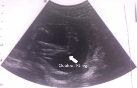
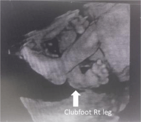
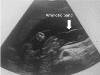
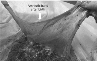
A Thai woman aged 30 years, G2P1A0L1, with gestational age of 21 weeks came to receive first antenatal care when her gestational age was 8+5 weeks and met her obstetrician on regular appointment. She had no congenital disease and history of illness with genetic diseases as well as refused the use of addictive drugs or substances, including no previous surgery. Her serological laboratory results were normal, last menstrual period (LMP) was 20/6/2018, and expected date of delivery (EDD) was 3/4/2019. After general pregnancy examination, it was found that her uterine size was correlated with gestational age. The obstetrician performed an ultrasound examination to assess the health of the fetus in accordance with the criteria of the antenatal department and found that the fetus’s right foot twisted inwards (Figures 1,2) and there was a large membrane located at the amnion tissues floating in the amniotic fluid (Figures 3,4). However, no abnormality of other organs of the fetus was found. The fetus’s weight was approximately 418 grams. Pregnant women was sent to referral hospital for second opinion which confirmed our findings , i. e. the fetus’ s right foot twisted inwards and there was a large band located at the amnion tissues floating in the amniotic fluid. Therefore, the hospital gave advice and sent her back to receive antenatal care at the original hospital. Because the membrane did not bind other organs, there was no need for antenatal treatment with surgery to cut it off. She had an appointment to meet her obstetrician every 3 weeks.
When her gestational age reached 24 weeks, the pregnancy examination showed that her uterine size was correlated with gestational age. According to the ultrasound examination result, there was a large amniotic band located at the amnion tissues floating in the amniotic fluid, the fetus’ s right foot twisted inwards, and other organs of the fetus were not tied with such membrane. She came to the hospital with labor pain at gestational age of 38+1 weeks with 11 times of antenatal care. There were no abnormalities found during pregnancy and had normal vaginal delivery. The newborn was female with birth weight of 3,500 grams, Apgar score of 9-10-10, and normal general appearance. The newborn’s right foot twisted inwards and the obstetrician diagnosed it as right clubfoot. Finally, the newborn was sent to a larger regional hospital with a pediatric surgeon to continue treatment of this abnormality in the post-natal period.
Discussion
Amniotic band syndrome is rare and unpreventable, but it is a major cause of birth defects in the fetus. The severity depends on the size of the amniotic band and the position of the fetus’s body being bound.
A two- or three-dimensional ultrasound examination will help diagnose severe disorders or fetal disabilities quickly and accurately
In general, the obstetrician will be able to diagnose it before childbirth in the second trimester of pregnancy with two- or three-dimensional ultrasound examination. If a fetus has a complicated disability, the obstetrician may consider giving advice on abortion to pregnant women [10]. Amniotic band syndrome is believed to be caused by rupture of fetal tissues in the amnion layer or chromosomal abnormalities. As a result, membranes binding fetal tissues float in the amniotic fluid and bind the fetus’s organs. When moving or wriggling [11], the fetus may have birth defects, such as bent or strained arms, legs or fingers [12], no skull, deformed face, cleft lip, cleft palate, nose twisted into the body, small and asymmetric eyes, cleft breasts, bent spine, and cleft abdominal wall [13].
Treatment of amniotic band syndrome that affects the fetus may be conducted by cutting off the membranes binding around the organs in some cases, but there is a risk of abortion. If a fetus is severe abnormalities due to such membranes and it is detected during the second trimester of pregnancy, the obstetrician may consider terminating the pregnancy. However, if there are membranes but they do not bind any organ, the pregnancy is mostly allowed to continue until maturity along with providing mother’s mental care because the mothers will have high anxiety, which will result in stress in the fetus as well. After the fetus is born, the treatment will be then performed, such as by wearing special shoes or doing surgery to adjust the structure of the feet [14].
Conclusion
Amniotic band syndrome is a rare and unpreventable syndrome. It is believed to be caused by rupture of fetal tissues in the amnion layer or chromosomal abnormalities. As a result, amniotic band binding fetal tissues float in the amniotic fluid and bind the fetus’s organs when the fetus is moving or wriggling. The severity depends on the fetus’s organs being bound. In most cases, it occurs at the fetus’s limbs, fingers, and toes, including other organs, such as deformed skull, face, mouth, nose, eyes, chest, ribs, spinal bone, front surface of sexual organs, and anal hole, which can result in the fetus’ s congenital malformation. A two- or three- dimensional ultrasound examination will help diagnose severe disorders or fetal disabilities quickly and accurately. If it is detected during the second trimester of pregnancy, the obstetrician may consider terminating the pregnancy. However, if there are membranes but they do not bind any organ, the pregnancy is mostly allowed to continue until maturity along with providing mother’s mental care because the mothers will have high anxiety, which will result in stress in the fetus as well. After the fetus is born, the treatment will be then performed, such as by wearing special shoes or doing surgery to adjust the structure of the feet.
- Cignini P, Giorlandino C, Padula F, Dugo N, Cafa EV (2012) Epidemiology and risk factors of amniotic band syndrome, or ADAM sequence. J Prenat Med 6: 59-63. Link: http://bit.ly/339zHgv
- Torpin R (1965) Amniochorionic Mesoblastic Fibrous Strings and Amnionic Bands: Associated Constricting Fetal Malformations or Fetal Death. Am J Obstet Gynecol 91: 65-75. Link: http://bit.ly/2yyqG2I
- Froster UG, Baird PA (1993) Amniotic band sequence and limb defects: data from a population-based study. Am J Med Genet 46: 497-500. Link: http://bit.ly/2KcKLlS
- Mukherjee PM, Natera M, Drew H, Creanga A (2019) Amniotic Band Syndrome: A Multidisciplinary Care Approach to the Treatment of a Rare Case. Cleft Palate Craniofac J 56: 105-109. Link: http://bit.ly/2GKmIID
- Ashutosh Halder (2010) Amniotic band syndrome and/or limb body wall complex: split or lump. Appl Clin Genet 3: 7-15. Link: http://bit.ly/2ZtE4AO
- Coady MSE, Moore MH, Wallis K (1998) Amniotic band syndrome: the association between rare facial clefts and limb ring constrictions. Plast Reconstr Surg 101: 640-649. Link: http://bit.ly/2YGMMuB
- Turğal M, Ozyüncü O, Yazıcıoğlu A, Onderoğlu LS (2014) Integration of three-dimensional ultrasonography in the prenatal diagnosis of amniotic band syndrome: A case report. J Turk Ger Gynecol Assoc 15: 56-9. Link: http://bit.ly/2LVXw6g
- Byrne J, Blanc WA, Baker D (1982) Amniotic band syndrome in early fetal life. Birth Defects Orig Artic Ser 18: 43-58. Link: http://bit.ly/336RhC4
- Kavita Hotwani, Krishna Sharma (2015) Oral rehabilitation for amniotic band syndrome: an unusual presentation. Int J Clin Pediatr Dent 8: 55-57. Link: http://bit.ly/2KeE2rz
- Ferris NJ, Tien RD (1994) Amnion Rupture Sequence with 'Exencephaly': MR Findings in a Surviving Infant .American Journal of Neuroradiology 15: 1030-1033. Link: http://bit.ly/2LVKCoU
- Goldfarb CA, Sathienkijkanchai A, Robin NH (2009) Amniotic constriction band: a multidisciplinary assessment of etiology and clinical presentation. J Bone Joint Surg Am 91: 68-75. Link: http://bit.ly/2GJ6Mq0
- Sunil Richardson, Rakshit Vijay Khandeparker, Philippe Pellerin (2017) Amniotic constriction band: a report of two cases with unique clinical presentations. J Korean Assoc Oral Maxillofac Surg 43: 171-177. Link: http://bit.ly/2yyp3Sp
- Anthony LH Moss, Tamsin A Burgues (2015) Monochorionic, Diamniotic Twins with Amniotic Band Syndrome at Exactly the Same Site for each: A Rare Amniotic Band Syndrome Presentation. Journal of Clinical Neonatology 4: 54-56. Link: http://bit.ly/2OK7KsO
- Anitha Ananthan, Gayatri Athalye Jape, Jean Du Plessis, Peter Annear, Rohan Page, et al. (2017) Retraction Notice to: Amniotic Band Syndrome with Pseudoarthrosis of Tibia and Fibula: A Case Report. The Journal of Foot & Ankle Surgery. 56:1121-1124. Link: http://bit.ly/2YBQglY

Article Alerts
Subscribe to our articles alerts and stay tuned.
 This work is licensed under a Creative Commons Attribution 4.0 International License.


 Save to Mendeley
