Global Journal of Medical and Clinical Case Reports
Neurological Outcome in a Term Infant with Tentorial Laceration Leading to Subdural Haemorrhage Following Vacuum-Assisted Delivery: A Case Report and Literature Review
Hee Chan Kang1 and Suresh Chandran2*
2Senior Consultant, Department of Neonatology, KK Women’s and Children’s Hospital, Singapore.
Adjunct Asst Professor Duke-NUS Medical School, Yong Loo Lin School of Medicine, Lee Kong Chian School of Medicine, Singapore
Cite this as
Chan KH, Chandran S (2016) Neurological Outcome in a Term Infant with Tentorial Laceration Leading to Subdural Haemorrhage Following Vacuum-Assisted Delivery: A Case Report and Literature Review. Glob J Medical Clin Case Rep 3(1): 035-037. DOI: 10.17352/2455-5282.000031There is a paucity of literature on this potentially devastating side effect of vacuum-assisted delivery. We present here a significant subdural haemorrhage associated with tentorial tear following vacuum-assisted delivery that resolved with only supportive medical measures without resulting in any neurological deficits nor delays on thorough examination and formal neuro-developmental assessment.
Introduction
About 8% of women in the general population are quoted to experience prolonged labour [1]. Simpson first described vacuum extraction in 1706 to a patient in labour for four days using “a cupping glass fixed to the scalp with an air pump” [2]. With growing concerns surrounding the “tedious and trying cases” of prolonged second stage of labour, the “Air Tractor” was the first publically introduced and used for vacuum-assisted delivery, attributed to Simpson in 1848 [2,3]. Interestingly one of his students, Mitchell claimed that this idea was stolen from him [4,5] with several attempts to take back the ownership in writing [5,6]. As Simpson would have hoped for, technological progression allowed modern devices using the same principle of vacuum force to become arguably the most successful instrument in alleviating the burdens of prolonged labour [7]. Unfortunately one of the potentially catastrophic adverse effects from its use is neonatal subdural haemorrhage; some requiring invasive surgery to alleviate the intracranial pressure resulting from this. Only sporadic case reports exist in the literature regarding this matter thus neither a pattern in outcomes nor perhaps guidelines to minimise this risk has not yet been established. This case report therefore aims to contribute to the literature pool the outcome and its implications on management of this clinical problem.
Case Report
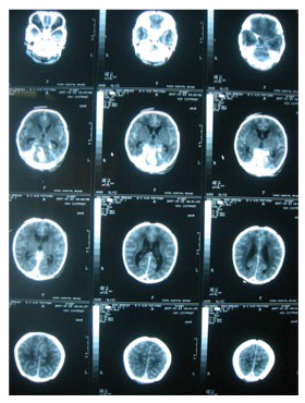
A 3280gm male was born to a 21 year-old gravida 3 para 2 mother with gestational diabetes on insulin; requiring vacuum-extraction due to poor progress. The previous 2 children were born via normal vaginal delivery and birthing process was uneventful. No known family history of bleeding diathesis. Feeding intolerance was noted but at 5 hours of life, the baby developed profound generalised seizures that required loading with phenobarbitone in addition to bolus doses of lorazepam. A bedside cranial ultrasound (CUS) was performed that demonstrated posterior fossa haemorrhage. He was intubated due to profound apnea and required positive pressure ventilation for 2 days. Once stabilised, a cranial computed tomography (CT) scan was done and confirmed infratentorial subdural haemorrhage (Figure 1). His coagulation profile, haematological and biochemical parameters were unremarkable. He made a uneventful recovery over 5 days and was discharged home on day 10 of life. He was on regular follow-up until 2 years of age. He acquired his mile stones at appropriate ages and had a formal Bayley-ll neuro-developmental assessment at 2 years of age, which was well within the normal range.
Discussion
The Rule of 4’s
Four main types of instrument-assisted deliveries are clearly discussed in a systematic review on forceps (rigid or soft), metal-cup, soft-cup and the handheld (which has a manual vacuum pump rather than an automated device) ventouse [8]. The reviewers concluded that whilst the forceps and metal-cup were associated with better chances of achieving successful vaginal delivery, they also carried increased risk of trauma to the mother and the new born. In this way obstetricians may be inclined to apply vaccum assisted delivery (VAD) with the metal-cup over the forceps, which has similarly high rates of successful vaginal delivery but less traumatic to the mother. Metal-cup ventouse resulted in more frequent scalp injuries and cephalhaematoma than its soft-cup counterparts, which had higher failure rates due to reduced traction force achieved [9]. The consequences of subdural haemorrhage was not addressed at length in this particular review probably in part due to the fact that its true incidence following VAD cannot be determined. Subdural haemorrhage (SDH) can occur even in uncomplicated vaginal deliveries of asymptomatic term infants [10], up to 45.5% detected by MRI brain is reported in one study [11].
Four steps to identify neonatal intra-cranial haemorrhage were outlined by Volpe in 1987 [12], assess for risk factors, observe closely for clinical signs, cerebrospinal fluid (CSF) extraction and diagnostic imaging. There is a slight male preponderance perhaps attributed to their greater occipito-frontal circumference; thus making them more vulnerable to vertical traction forces generated during the process [13]. Risk factors for developing SDH in general are outlined in Table 1 [12]. Clues in the neonate can be highly variable and include any number or combination of the following; bulging fontanellae, increased occipito-frontal circumference, apnoea, bradycardia, respiratory distress, retinal haemorrhage, lethargy, fever, opisthotonus and seizures [14,15]. Volpe inferred in an article that slow-reacting or unreactive pupils due to compression of the occulomotor nerve are the most distinctive sign of posterior fossa [12]. Apnoea or seizures were the two most common presentation in a series and such symptoms following vacuum extraction should raise suspicion [15] as seen in our case. In Huang et al case series, heavily blood-stained CSF was found in all 6 cases in whom lumbar puncture was possible [13]. On analysis of CSF, typical characteristics include high red blood cell count, raised protein, mildly raised lymphocytes and low glucose, although hypoglycorrhachia is said to occur only between 5-15 days from the onset of intracranial haemorrhage. It is almost uniformly associated with concurrent low lactate levels, but the mechanism of this phenomenon is unclear [12]. CUS and CT brain are the mainstay of imaging. Although significant bleeds in the posterior fossa can be picked up by CUS, as noted in our case, being and operator-dependent procedure, it has been deemed rather insensitive, particularly for retrocerebellar bleeds unless large enough to cause cerebellar displacement [13]. CT scan is therefore advised for confirmation [14] and thus the initial management would depend on clinical judgment until then. Blauwblomme et al.. Concluded that MRI in the acute setting did not influence their decision for surgery but suggested that later scans may help with the prognosis [15].
Four major types of intra-cranial haemorrhage seen in the neonate are (extra-dural is also associated with VAD but rare [16] and therefore excluded for the purpose of this discussion); subdural, subarachnoid, intracerebellar and periventricular-intraventricular haemorrhage. The majority of such cases are attributed to parturitional trauma. The subdural type is the only one more commonly associated with term infants; the rest are with premature counterparts [12]. The main mechanism of injury relates to foetal head moulding whereby the antero-posterior axis lengthens and the biparietal axis shortens [17].
Four major sources of SDH exist and are outlined in Table 2 [12]. Tentorial laceration is the most commonly reported injury seen in vacuum extraction due its anatomy vulnerable to vertical traction forces. The vertical parts of the tentorium cerebelli (running up beside the straight sinus, later fusing to becoming the falx cerebri) is under the greatest strain when the foetal head is forced to assume brachycephalic and turricephalic shapes during labour [15,18]. This transient moulding of the head shape also causes stretching of the internal cerebral veins, basal veins of Rosenthal, the vein of Galen, the infratentorial veins, lateral and straight sinuses [12,15]. The vein of Galen appears to be even less adaptable to the vertical shear force as its distal end is attached to the dura at the straight sinus whilst its proximal is relatively free to move [15,18].
Management
Management should be tailored to the individual baby in a multi-disciplinary approach. It is summarised well by Blauwblomme et al. [15]. Supportive medical therapy in suppressing the seizures if present and treatment as per guidelines for raised intracranial pressure (ICP) and cerebral oedema should be followed. If signs of raised ICP persist despite treatment, associated abnormal physiology must be ruled out/corrected; hypercarbia, hypoxaemia, hypotension, hypoglycaemia, pain and seizures [19]. The patient must be monitored very closely for any signs of persistent or worsening neurological status at which surgical intervention must be considered; particularly if there is any evidence of brain stem dysfunction or obstructive hydrocephalus [15]. Coagulopathy should be corrected if present.
Conclusion
Therefore two conclusions arose from this case; though uncommon, such intracranial injuries can arise despite the long history of use of VAD and its application by experienced hands; secondly, a careful observation with prompt supportive medical therapy can prevent long-term neurological deficit without surgical intervention. We hope to increase awareness amongst neonatologists in identifying and therefore treating such neonates swiftly.
- Nystedt, A, Hildingsson I (2014) Diverse definitions of prolonged labour and its consequences with sometimes subsequent inappropriate treatment. BMC Pregnancy Childbirth 14: 233. Link: https://goo.gl/nnerUv
- Kozak LJ, Weeks JD (2002) U.S. trends in obstetric procedures, 1990-2000. Birth 29: 157-161. Link: https://goo.gl/WTLC2c
- Simpson JY (1849) The Air Tractor as a Substitute for the Forceps in Tedious Labors (Edinburgh Monthly Journal of Medical Sciences 556.
- Strickland RA (2008) James Y Simpson: a physician ahead of his time. Bull Anesth Hist 26: 6-7. Link: https://goo.gl/cazYVl
- Mitchell J (1849) The air-tractor. Lancet 1: 491-492.
- Mitchell J, Simpson JY (1849) Who invented the air-tractor? Correspondence between Dr. Simpson and Dr. Mitchell. London Medical Gazette 8: 519-521.
- Dexeus JM, Ruiz JG (1963) 3 cases of grave fetal accidents secondary to the use of the vaccum extractor. Rev Esp Obstet Ginecol 22: 440-444. Link: https://goo.gl/2miDje
- O'Mahony F, Hofmeyr GJ, Menon V (2010) Choice of instruments for assisted vaginal delivery. Cochrane Database Syst Rev Cd005455. Link: https://goo.gl/YrShBi
- Hofmeyr GJ, Gobetz L, Sonnendecker EW,Turner MJ (1990) New design rigid and soft vacuum extractor cups: a preliminary comparison of traction forces. Br J Obstet Gynaecol 97: 681- 685. Link: https://goo.gl/TAZ8yv
- Whitby EH, Griffiths PD, Rutter S, Smith MF, et al. (2004) Frequency and natural history of subdural haemorrhages in babies and relation to obstetric factors. Lancet 363: 846-851. Link: https://goo.gl/nigl8Y
- Rooks VJ, Eaton JP, Ruess L, Petermann GW, Keck-Wherley J, et al. (2008) Prevalence and evolution of intracranial hemorrhage in asymptomatic term infants. AJNR Am J Neuroradiol 29: 1082-1089. Link: https://goo.gl/0KYk91
- Volpe JJ (1987) Intracranial haemorrhage, Chapter 10, Neurology of the Newborn, 2nd ed. 282-294.
- Huang CC, Shen EY (1991) Tentorial subdural hemorrhage in term newborns: Ultrasonographic diagnosis and clinical correlates. Pediatr Neurol 7 171-177. Link: https://goo.gl/2W02Kh
- Brouwer AJ, Groenendaal F, Koopman C, Nievelstein RJA, Han SK, et al. (2010) Intracranial haemorrhage in full-term newborns: a hospital-based cohort study. Neuroradiology 52: 567- 576. Link: https://goo.gl/3dRFpc
- Blauwblomme T, Garnett M, Vergnaud E, Boddaert N, Bourgeois M, et al. (2013) The management of birth-related posterior fossa hematomas in neonates. Neurosurgery 72: 755- 762. Link: https://goo.gl/nUHLDh
- Noguchi M, Inamasu J, Kawai F, Kato E, Kuramae T, et al. (2010) Ultrasound-guided needle aspiration of epidural hematoma in a neonate after vacuum-assisted delivery. Childs Nerv Syst 26: 713-716. Link: https://goo.gl/ZDW4cf
- Lapeer RJ, Prager RW (2001) Fetal head moulding: finite element analysis of a fetal skull subjected to uterine pressures during the first stage of labour. J Biomech 34: 1125-1133. Link: https://goo.gl/juOF7b
- Harpold TL, McComb JG, Levy ML (1998) Neonatal neurosurgical trauma. Neurosurg Clin N Am 9: 141-154. Link: https://goo.gl/nTCPeF
- Kliegman R, Nelson WE (2011) Nelson textbook of pediatrics (19th ed.). Philadelphia, PA: Elsevier/Saunders.

Article Alerts
Subscribe to our articles alerts and stay tuned.
 This work is licensed under a Creative Commons Attribution 4.0 International License.

 Save to Mendeley
